Access Menu
What is Deep Vein Thrombosis (DVT)?
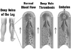
Deep vein thrombosis is the formation of a blood clot in a deep leg vein. It is a very serious condition that can cause death or permanent damage to the leg. In the United States alone, 600,000 people with DVT are admitted to hospitals each year. One in every 100 DVT patients dies.
How does a DVT form?
The deep leg veins are surrounded by powerful muscles that contract to force blood upward against gravity, back to the lungs and heart. One-way valves inside the veins prevent downward back-flow of blood. Blood clots form when the body’s clotting system functions abnormally, and when the circulation of blood slows down due to illness, injury or inactivity.
What are the risk factors for DVT?
Factors that increase your risk for DVT include:
- Previous DVT or family history of DVT
- Prolonged immobility, which can be due to stroke, paralysis, prolonged bedrest or sitting, or prolonged air travel (known as “economy class syndrome”)
- Recent surgery
- Hormone therapy or oral contraceptives
- Current or recent pregnancy
- Trauma and/or orthopedic procedures
- Varicose veins
- Congestive heart failure
- Use of central venous catheters
- Inflammatory bowel disease
- Inherited blood clotting (coagulation) abnormalities
- Obesity
- Age above 40 years
What are the typical symptoms of DVT?
Common symptoms include:
- Previous DVT or family history of DVT
- Prolonged immobility, which can be due to stroke, paralysis, prolonged bedrest or sitting, or prolonged air travel (known as “economy class syndrome”)
- Swelling of the leg
- Calf or leg pain or tenderness
- Leg fatigue
- Discoloration of the legs
- Surface veins become more visible
How is a DVT diagnosed?
If you suspect that you may have a DVT, you should immediately go to an emergency room to be evaluated by a physician – it’s a life threatening disease. If your physician agrees that DVT is a possibility, he/she will probably order an ultrasound examination of your leg veins. In some cases, DVT is diagnosed by an Interventional Radiologist using a venogram, which is an X-ray image of your veins – this test allows the physician to see inside your veins and locate a clot that is blocking blood flow. Interventional Radiologists can not only identify the blood clot, but they can also often eliminate the clot and treat underlying problems in the veins that may have caused the blood clot to form.
What problems does DVT cause?
Pulmonary Embolism
When DVT is left untreated, a piece of clot can break off and travel through circulation to the lungs – this is known as pulmonary embolism. The clot can interfere with the lung’s ability to provide oxygen to the body, and when severe, this can cause heart failure and/or death. With early treatment using blood-thinners, people with DVT can reduce their chances of developing a life-threatening pulmonary embolism to less than 1 percent.
Post-thrombotic Syndrome
Post-thrombotic syndrome is an underrecognized but common long-term consequence of DVT. When DVT is treated with blood thinners alone, the blood clot is not actively dissolved. Rather, the blood thinners prevent new clots from forming, but the already present clot remains in the leg. The body may eventually fully or partially dissolve the clot, but the vein valves often become damaged in the meantime. The continued vein blockage and valve damage cause abnormal pooling of blood in the leg, which can cause the post-thrombotic syndrome. Patients with the post-thrombotic syndrome often experience chronic leg pain or heaviness, swelling, difficulty walking, changes in skin color and texture, and in severe cases, skin ulcers (sores). These symptoms may develop within several months or over years. Unfortunately, these problems occur to some degree in as many as 60-70 percent of people with DVT.
How is a DVT treated?
DVT is treated with blood thinners to prevent pulmonary embolism and to prevent further clot formation. In patients with extensive DVT, an additional treatment option is catheter-directed thrombolysis. In patients who cannot receive blood thinners or in whom blood thinners have failed to prevent further clot formation, an additional treatment option is the placement of an inferior vena cava filter.
Catheter-Directed Thrombolysis
Catheter-directed thrombolysis is performed under imaging guidance by Interventional Radiologists. This procedure is designed to rapidly break up the clot, restore blood flow within the vein, and potentially preserve valve function to minimize the risk and severity of post-thrombotic syndrome. Using imaging guidance, the Interventional Radiologist inserts a catheter into a leg vein behind the knee and threads it into the vein containing the clot. A clot-dissolving drug is infused through the catheter directly into the clot and repeated over one to two days. The fresher the clot, the faster it dissolves. A venogram is repeated to demonstrate that the clot has dissolved, and to detect any narrowing in the vein that might lead to future clot formation. If vein narrowing is present, the Interventional Radiologist can treat it using balloon angioplasty or by placing a stent.
Vena Cava Filter
In patients in whom catheter-directed thrombolysis and blood thinners are not medically appropriate, an Interventional Radiologist can insert a small filtering device into the large vein that drains the legs (the inferior vena cava) under imaging guidance. This vena cava “filter” functions like a catcher’s mitt to capture blood clots that break off.