Access Menu

Why all the fuss about cigarettes? People have been smoking for centuries.

It wasn’t until 1964, when the Surgeon General of the United States issued the first reports on the dangers of cigarette smoking, that there was clear, convincing evidence that smoking is dangerous. New studies continually add to the evidence, so today it can be said without question that smoking kills. Cigarettes alone kill more than 50,000 Canadians each year – more than alcohol, heroin, crack, automobile and airplane accidents, murders, suicides and AIDS combined.

How does tobacco actually hurt my body?

More than 4,000 chemicals are emitted by a lit cigarette – 43 of them are known to cause cancer. The three most dangerous chemicals from smoke are:
tar, which is capable of causing cancer in tissues it comes in contact with;
nicotine, a highly addictive drug that is absorbed by the lungs and affects the nervous system; and
carbon monoxide, a deadly gas that reduces the ability of your blood to carry oxygen throughout your body.
When you smoke, you increase the rate of your heartbeat and encourage the growth of plaque – a sticky substance that builds up in the arteries and causes hardening of the arteries which contributes to heart disease.
Cigarette smoke also destroys the tiny hairs that line the airways to your lungs, which leads to being more susceptible to lung infections and irritation.
The cancers that are most likely to result from smoking occur because the chemicals in the smoke come into direct contact with your tissues. For example, inhaling smoke brings the chemicals in contact with your mouth, throat and lungs. Smokers swallow a certain amount of smoke exposing the esophagus and digestive tract to the chemicals. The chemicals also are absorbed into the bloodstream and cleaned from the blood in the bladder. Cancers in all of these organs happen anywhere from twice as often to 14 times as often in smokers as in nonsmokers.

Smoking is my business. Why should anyone else care what I do?

Your smoking is a danger to innocent people, especially children. Second-hand smoke causes 150,000 to 300,000 lower respiratory tract infections, such as bronchitis and pneumonia, in young children each year. Of these, between 7,500 and 15,000 result in hospitalization. Plus, a pregnant woman can harm or kill her unborn child by smoking.

Am I safe having only three or four cigarettes a day?

Smoking in any form and in any amount is harmful to you. For example, smoking just two cigarettes a day doubles your risk of lung cancer. The number of cigarettes you smoke, the amount of smoke you take into your lungs and the length of time you smoke all count against you.

Seriously, what are the chances that I’m going to die from smoking?

One in five Americans die each year from tobacco. Smoking accounts for two of every 10 deaths from coronary heart disease or stroke, three of every 10 cancer deaths, and more than eight of every 10 deaths from chronic lung diseases such as emphysema and chronic bronchitis.
A. Even if it doesn’t kill you, smoking will complicate your life. If you smoke, you are more likely to have blockages of blood vessels to the kidneys, legs and brain. These blockages can lead to high blood pressure, pain with walking and stroke. You are also more likely to have complications from birth control pills, more likely to have a miscarriage, more likely to have coughs and colds, and more likely to have osteoporosis – a weakening of your bones.

If I quit now, will it improve my health?

Quitting does reduce the risk. Within five years your risk is cut in half; after 15 years, the risk has practically disappeared. That’s why we recommend you quit. Now.

What is an interventional radiologist?

Interventional radiologists are doctors who specialize in minimally invasive, targeted treatments performed using imaging for guidance. They use their expertise in reading X-rays, ultrasound, MRI and other diagnostic imaging equipment to guide tiny instruments, such as catheters, through blood vessels or through the skin to treat diseases without surgery. Interventional radiologists are board certified radiologists that are fellowship trained in nonsurgical interventions using imaging guidance. Many illnesses caused by smoking are treated by interventional radiologists.

Some of the diseases and conditions that are directly linked to smoking are:

A. aortic aneurysm, aortic atherosclerosis, arterial vascular disease, asthma, bladder cancer, bronchitis, cancer of the esophagus, cancer of the mouth and tongue, cervical cancer, chronic obstructive lung disease, coronary atherosclerosis, coronary heart disease, early menopause, emphysema, endometrial cancer, gastric cancer, sinfertility, kidney cancer, larynx cancer, liver cancer, lung cancer, osteoporosis, pancreatic cancer, peptic ulcer, stomach cancer, stroke, throat cancer

Exposure to someone else’s smoke (secondhand smoke) has been directly linked to these conditions in children:

Allergy, asthma, bronchitis, cleft lip, cleft palate, congenital heart disease, coughs, cystic fibrosis, dizziness, ear diseases, headaches, hernia, hoarseness, itchy nose, lower height, phlegm, pneumonia, premature birth, reduced lung function, runny nose, sneezing, snoring, sore eyes, sore throats, sudden infant death syndrome, tonsillitis, upper respiratory tract infections, upset stomach, viral respiratory infection, wheezing

Pregnant women who smoke endanger their unborn children, who can suffer from the following conditions:

Congenital malformations, damage to the placenta, damage to umbilical blood vessels, enlarged placenta, fetal and infant mortality, increased incidence of premature birth, low birthweight, neonatal deaths, spinal malformation, spontaneous abortion, stillbirth, sudden infant death syndrome, tubal pregnancy, urogenital abnormalities

Access Menu

What is a stent?

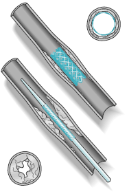
A stent – sometimes called an endoprosthesis – is a small, flexible tube made of medical grade plastic or wire mesh. It is implanted in the body to treat a variety of medical conditions.

What conditions can be treated with stents?

Stents are used to treat a number of medical conditions:

Stents are most commonly used to hold clogged blood vessels open after angioplasty, a procedure in which a balloon on the end of a catheter is moved through the body to the site where the blood vessel is blocked. The balloon is then inflated to open the vessel. In some cases, however, stents may be placed as the primary means for holding the vessel open. Stents also are used to hold open bile ducts or other pathways in the body that have been narrowed or blocked by tumors or other obstructions. Areas where stents are most often used for this reason include:
  • the esophagus, to treat blockages or narrowings that make it difficult to swallow;
  • the bile ducts in the pancreas or liver, when an obstruction prevents bile from draining into the digestive tract; and
  • the airways of the lungs, to treat obstructions that interfere with normal breathing.
In some cases, stents are used to treat patients with severe liver disease. These patients often experience life-threatening bleeding because their livers are too damaged to handle the normal flow of blood. One way to treat this condition is with a technique called TIPS, in which a stent is used to create a connection between two veins and divert the flow of blood away from the liver.

How are stents implanted in the body?

The interventional radiologist makes a very small incision in the skin, about the size of a pencil tip. The stent, which is placed on the end of a catheter, is threaded under X-ray guidance to the area of treatment. This interventional radiology technique is generally less traumatic for the patient than surgical implantation because it involves smaller incisions, less pain and shorter hospital stays.

Access Menu

What is TIPS?

TIPS is a medical procedure that helps correct blood flow problems in the liver, which is a common side effect of liver disease. The procedure is done without surgery. It is performed in the radiology department by a specially trained doctor known as an interventional radiologist.

Why would a person need TIPS?

TIPS can often help a condition called portal hypertension, which occurs when the normal flow of blood through the blood vessels in the liver is slowed or blocked by scarring or other damage. The scarring or damage is caused by liver diseases, such as hepatitis or cirrhosis.
Several things can happen when the blood vessels are blocked. Sometimes bleeding occurs when the blood tries to find unblocked pathways. In other instances, fluid accumulates in the area around the stomach. (This is called ascites.) Sometimes fluid accumulates in the chest. (This is called pleural fluid).

What happens during TIPS?

The interventional radiologist will make a tunnel in the liver through which the blocked blood can flow. After the tunnel is made, the doctor will insert a small metal tube (called a shunt or stent) into the tunnel to make sure the tunnel stays open.
The interventional radiologist will use x-ray pictures and x-ray dye (also called contrast) to guide the procedure.

How do I prepare for the procedure?

Do not eat or drink after midnight. If you are allergic to x-ray dye (contrast) or iodine, let your doctor know as soon as possible so that special precautions can be taken during the procedure.
Before the procedure, a member of the interventional radiology team will talk with you about the procedure in detail and answer any questions you have.

What is TIPS like? Will it hurt?

Patients usually receive medication before TIPS to make them very groggy during the procedure. Sometimes patients are put completely to sleep by an anesthesiologist. In any case, the patient will be given medication to take away pain.
The interventional radiologist performs the entire procedure through an I.V. tube in the neck. If you are not put completely to sleep, the doctor will numb the area before putting in the I.V. tube.
When the procedure is finished, the doctor may leave the I.V. tube in your neck so you can receive other medication through it. Stitches are not necessary when the I.V. tube is removed.

What happens after my TIPS?

You will return from the radiology department to your room (or possibly to an intensive care unit, if your doctor wants to keep you under close observation). The doctor and nurses will tell you when you can eat and how long you will have to stay in bed.
Your stomach and/or neck may be sore. There also may be some swelling or bruising in your neck. The soreness and swelling will go away in a few days to a week and generally requires no treatment.
Your TIPS will require periodic check-ups. Your doctor will need to make sure that the TIPS is working well. This is usually done at three, six, or 12 months after the TIPS procedure. Your doctor will tell you the exact schedule you should follow.

What are the risks of TIPS?

Any procedure involving blood vessels raises the risk of bleeding. Recent studies indicate that the risk of serious bleeding is about one to two percent.
Some patients get sick from the x-ray dye. This is more likely in patients with diabetes, kidney disease, asthma or a previous allergic reaction to x-ray dye. If you have one of these conditions, tell your interventional radiologist before the procedure begins.
About ten percent of the time, patients become temporarily confused or disoriented as a result of TIPS. This can usually be treated with medication after the TIPS procedure.
Because everyone is different, there may be risks that are not mentioned here. The risks to you will be discussed in more detail by a member of your interventional radiology team.

Are there alternatives to TIPS?

The blockage of blood flow through the liver is sometimes best treated with standard surgery and sometimes best treated with TIPS. Your doctors will tell you which they think is the best option for you.

What are the benefits of TIPS?

The benefit of TIPS is that your symptoms can be relieved without surgery, and the recovery time is less than it would be after surgery.

Access Menu


What are uterine fibroids?

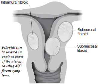
Fibroid tumors are noncancerous (benign) growths that develop in the muscular wall of the uterus. While fibroids do not always cause symptoms, their size and location can lead to problems for some women, including pain and heavy bleeding. They typically improve after menopause when the level of estrogen, the female hormone that circulates in the blood, decreases dramatically. However, menopausal women who are taking supplemental estrogen (hormone replacement therapy) may not experience relief of symptoms. Fibroids range in size from very tiny to the size of a cantaloupe or larger. In some cases they can cause the uterus to grow to the size of a five-month pregnancy or more. Fibroids may be located in various parts of the uterus.
There are three primary types of uterine fibroids:
  • Subserosal fibroids, which develop in the outer portion of the uterus and expand outward. They typically do not affect a woman’s menstrual flow, but can become uncomfortable because of their size and the pressure they cause.
  • Intramural fibroids, which develop within the uterine wall and expand, making the uterus feel larger than normal. These are the most common fibroids. This can result in heavier menstrual flows and pelvic pain or pressure.
  • Submucosal fibroids are deep within the uterus, just under the lining of the uterine cavity. These are the least common fibroids, but they often cause symptoms, including very heavy and prolonged periods.
You might hear fibroids referred to by other names, including myoma, leiomyoma, leiomyomata and fibromyoma.

What are typical symptoms?

Depending on location, size and number of fibroids, they may cause:
  • Heavy, prolonged menstrual periods and unusual monthly bleeding, sometimes with clots. This often leads to anemia.
  • Pelvic pain
  • Pelvic pressure or heaviness
  • Pain in the back or legs
  • Pain during sexual intercourse
  • Bladder pressure leading to a constant urge to urinate
  • Pressure on the bowel, leading to constipation and bloating
  • Abnormally enlarged abdomen

Who is most likely to have uterine fibroids?

Uterine fibroids are very common, although often they are very small and cause no problems. From 20 to 40 percent of women age 35 and older have uterine fibroids of a significant size. African-American women are at higher risk for fibroids: as many as 50 percent have fibroids of a significant size.

How are uterine fibroids diagnosed?

Fibroids are usually diagnosed during a gynecologic internal examination. Your doctor will conduct a pelvic exam to feel if your uterus is enlarged. The presence of fibroids is most often confirmed by an abdominal ultrasound. Fibroids also can be confirmed using magnetic resonance (MR) and computed tomography (CT) imaging techniques. Ultrasound, MR and CT are painless diagnostic tests. Appropriate treatment depends on the size and location of the fibroids, as well as the severity of symptoms.

How are uterine fibroids treated?

Most fibroids do not cause symptoms and are not treated. When they do cause symptoms, drug therapy often is the first step in the treatment. This might include a prescription for birth-control pills or other hormonal therapy, or the use of non-steroidal anti-inflammatory drugs, such as ibuprofen or naproxen sodium. In many patients, symptoms are controlled with these treatments and no other therapy is required. Some hormone therapies do have side effects and other risks when used long-term so they are generally used temporarily. Fibroids often grow back after therapy is discontinued. The next step is to try more invasive therapy.
The most common treatment options include:

Uterine artery (or fibroid) embolization

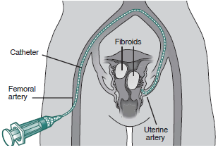
An interventional radiologist makes a tiny incision in the groin and passes a small tube called a catheter through the artery. When the catheter reaches the uterine artery, the interventional radiologist slowly releases tiny plastic particles the size of grains of sand into the vessels. The particles flow to the fibroids first and wedge into the vessels and cannot travel to other parts of the body. This blocks the blood flow to the tumor, causing it to shrink.

Myomectomy

Myomectomy is a surgical procedure that removes visible fibroids from the uterine wall. Myomectomy, like UFE, leaves the uterus in place and may, therefore, preserve the woman’s ability to have children. There are several ways to perform myomectomy, including hysteroscopic myomectomy, laparoscopic myomectomy and abdominal myomectomy:

  • Hysteroscopic Myomectomy: Hysteroscopic myomectomy is used only for fibroids that are just under the lining of the uterus and that protrude into the uterine cavity. There is no need for a surgical incision. The doctor inserts a flexible scope (hysteroscope) into the uterus through the vagina and cervix and removes the fibroids using special surgical tools fitted to the scope. Usually this is an outpatient procedure performed while the patient is under anesthesia and not conscious.
  • Laparoscopic Myomectomy: Laparoscopic myomectomy may be used if the fibroid is on the outside of the uterus. Small incisions are made so the doctor can insert a probe with a tiny camera attached and another probe fitted with surgical instruments inside the abdominal cavity and remove the tumors. It is performed while the patient is under general anesthesia and not conscious. The average recovery time is about two weeks.
  • Abdominal Myomectomy: This is a surgical procedure in which an incision is made in the abdomen to access the uterus, and another incision is made in the uterus to remove the tumor. Once the fibroids are removed, the uterus is stitched closed. The patient is given general anesthesia and is not conscious for this procedure, which requires a several-day hospital stay.

While myomectomy is frequently successful in controlling symptoms, the more fibroids there are in a patient’s uterus, generally, the less successful the surgery. In addition, fibroids may grow back several years after myomectomy.

Hysterectomy

Approximately one-third of the more than half-million hysterectomies performed in the United States each year are due to fibroids. In a hysterectomy, the uterus is removed in an open surgical procedure. This operation is considered major surgery and is performed while the patient is under general anesthesia. It requires three to four days of hospitalization and the average recovery period is about six weeks. Some women are candidates for a newer, laparoscopic procedure. The recovery time for this procedure is considerably shorter. Hysterectomy is the most common current therapy for women who have fibroids. It is typically performed in women who have completed their childbearing years or who understand that after the procedure, they cannot become pregnant.


What is fibroid embolization?

It is a minimally invasive procedure, which means it requires only a tiny nick in the skin. It is performed while the patient is conscious but sedated – drowsy and feeling no pain.
Fibroid embolization is performed by an interventional radiologist, a physician who is specially trained to perform this and other minimally invasive procedures. The interventional radiologist makes a small nick in the skin (less than 1⁄4 of an inch) in the groin and inserts a catheter into an artery. The catheter is guided through the artery to the uterus while the interventional radiologist watches the progress of the procedure using a moving X-ray (fluoroscopy). The interventional radiologist injects tiny plastic particles the size of grains of sand into the artery that is supplying blood to the fibroid tumor. This cuts off the blood flow and causes the tumor (or tumors) to shrink. The artery on the other side of the uterus is then treated.
Fibroid embolization usually requires a hospital stay of one night. Pain-killing medications and drugs that control swelling typically are prescribed following the procedure to treat cramping and pain, which are common side effects. Fever is an occasional side effect, and is usually treated with acetaminophen. Many women resume light activities in a few days and the majority of women are able to return to normal activities within one week.
While embolization to treat uterine fibroids has been performed since 1995, embolization of the uterus is not new. It has been used successfully by interventional radiologists for 20 years to treat heavy bleeding after childbirth. The procedure is now available at hospitals and medical centers across the country.

How successful is the fibroid embolization procedure?

Studies show that 78 to 94 percent of women who have the procedure experience significant or total relief of heavy bleeding, pain and other symptoms. The procedure also is effective for multiple fibroids. Recurrence of treated fibroids is very rare. In one study in which patients were followed for six years, no fibroid that had been embolized regrew.

Are there risks associated with the treatment of fibroid tumors?

Fibroid embolization is considered to be very safe, however, there are some associated risks, as there are with almost any medical procedure. Most women experience moderate to severe pain and cramping in the first several hours following the procedure. Some experience nausea and fever. These symptoms can be controlled with appropriate medications. A small number of patients have experienced infection, which usually can be controlled with antibiotics. It also has been reported that there is a 1 percent chance of injury to the uterus, potentially leading to hysterectomy. A small number of patients have entered into menopause after embolization. This is more likely to occur if the woman is in her mid-forties or older, and is already nearing menopause. Myomectomy and hysterectomy also carry risks, including infection and bleeding leading to transfusion. Patients who undergo myomectomy may develop adhesions causing tissue and organs in the abdomen to fuse together, which can lead to infertility. In addition, the recovery time is much longer for abdominal myomectomy, generally one to two months. You should talk with your doctor about possible side effects of any procedure you may choose.

Is fibroid embolization an FDA-approved procedure?

The FDA does not regulate the practice of medicine, but it does approve devices and medications. All devices, equipment and medications used for fibroid embolization are approved by the FDA for use in people. Many women wonder about the safety of leaving plastic particles in the body. It is reassuring to know that the particles most commonly used in UFE have been available with FDA approval for over 20 years. During that time, they have been used in thousands of patients without long-term complications.

List of facilities

Contact the following facilities only if your physician has confirmed that your symptoms are due to uterine fibroids.

Access Menu

There are many variations of passages of Lorem Ipsum available, but the majority have suffered alteration in some form, by injected humour, or randomised words which don’t look even slightly believable. If you are going to use a passage of Lorem Ipsum, you need to be sure there isn’t anything embarrassing hidden in the middle of text. All the Lorem Ipsum generators on the Internet tend to repeat predefined chunks as necessary, making this the first true generator on the Internet. It uses a dictionary of over 200 Latin words, combined with a handful of model sentence structures, to generate Lorem Ipsum which looks reasonable. The generated Lorem Ipsum is therefore always free from repetition, injected humour, or non-characteristic words etc.

Access Menu

Contrary to popular belief, Lorem Ipsum is not simply random text. It has roots in a piece of classical Latin literature from 45 BC, making it over 2000 years old. Richard McClintock, a Latin professor at Hampden-Sydney College in Virginia, looked up one of the more obscure Latin words, consectetur, from a Lorem Ipsum passage, and going through the cites of the word in classical literature, discovered the undoubtable source. Lorem Ipsum comes from sections 1.10.32 and 1.10.33 of “de Finibus Bonorum et Malorum” (The Extremes of Good and Evil) by Cicero, written in 45 BC. This book is a treatise on the theory of ethics, very popular during the Renaissance. The first line of Lorem Ipsum, “Lorem ipsum dolor sit amet..”, comes from a line in section 1.10.32.

Contrary to popular belief, Lorem Ipsum is not simply random text. It has roots in a piece of classical Latin literature from 45 BC, making it over 2000 years old. Richard McClintock, a Latin professor at Hampden-Sydney College in Virginia, looked up one of the more obscure Latin words, consectetur, from a Lorem Ipsum passage, and going through the cites of the word in classical literature, discovered the undoubtable source. Lorem Ipsum comes from sections 1.10.32 and 1.10.33 of “de Finibus Bonorum et Malorum” (The Extremes of Good and Evil) by Cicero, written in 45 BC. This book is a treatise on the theory of ethics, very popular during the Renaissance. The first line of Lorem Ipsum, “Lorem ipsum dolor sit amet..”, comes from a line in section 1.10.32.

Contrary to popular belief, Lorem Ipsum is not simply random text. It has roots in a piece of classical Latin literature from 45 BC, making it over 2000 years old. Richard McClintock, a Latin professor at Hampden-Sydney College in Virginia, looked up one of the more obscure Latin words, consectetur, from a Lorem Ipsum passage, and going through the cites of the word in classical literature, discovered the undoubtable source. Lorem Ipsum comes from sections 1.10.32 and 1.10.33 of “de Finibus Bonorum et Malorum” (The Extremes of Good and Evil) by Cicero, written in 45 BC. This book is a treatise on the theory of ethics, very popular during the Renaissance. The first line of Lorem Ipsum, “Lorem ipsum dolor sit amet..”, comes from a line in section 1.10.32.