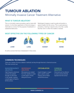
Access Menu


Tumour Ablation

Minimally Invasive Cancer Treatment Alternative

What is Tumour Ablation? 

This is a minimally invasive procedure used to treat solid cancers. It is the direct application of thermal energy or chemicals to a tumour for the purpose of eradication or substantial destruction. Radiological imaging is used to guide and position a special needle into the tumour. This is a percutaneous procedure, meaning it requires only a tiny hole, usually less than 3mm in which the needle is inserted.

Most Effective on the Following Types of Cancer:

  • Liver
  • Kidney
  • Lung
  • Bone
    • As well as cancers that have metastasized in these areas.  

Surgery or Ablation?

Patients that are not suitable for surgery can have tumour ablation as a safer alternative that can be either palliative or curative for their symptoms. Also, small tumours, can be effectively treated with tumour ablation preventing a much more invasive surgery. Ablation can dramatically shrink the size of a tumour, and for those that are 3cm or less in diameter has been an effective means of complete treatment, meaning that no residual cancer is present.

Common Techniques

There are a variety of tumour ablation methods, be they through thermal or chemical sources. The most common are:

  • Radiofrequency Ablation: high-frequency electrical currents are passed through an electrode in the needle, creating a small region of heat to kill the tumour
  • Microwave Ablation: microwaves are created from the needle to create a small region of heat killing the tumour
  • Cryoablation: liquid nitrogen or argon gas is used to create intense cold to freeze and kill the tumour
  • Chemical Ablation: chemicals such as ethanol or acetic acid are directly injected into the tumour

Access

A 2014 study looking at hospitals in Ontario identified that under 45% of hospitals offered Tumour Ablation, meanwhile 75% of hospitals would be willing to provide those services if the appropriate funding was provided.  The hospitals that did offer ablation procedures were focused in the Toronto area and in Southwest Ontario.

Benefits

  • Cancer treatment for patients who aren’t cleared for surgery
  • High level of success
  • Short recovery time
  • Proven results

Poster


List of facilities


Please note that the information contained on this page is intended solely for informational purposes and should not be used as a substitute for professional medical advice, diagnosis, or treatment. The list provided here includes only members of the Canadian Association for Interventional Radiology (CAIR) who have indicated that they perform the specified procedure. It is important to understand that this is not a comprehensive list of all physicians or facilities capable of performing this procedure.

The names, physicians, and facilities listed are members of CAIR and have provided their information voluntarily. While we strive to keep this information up to date and accurate, CAIR does not guarantee the accuracy, completeness, or timeliness of the information provided. The inclusion of any name in this list does not imply endorsement by CAIR or a recommendation of their services.

Patients are encouraged to conduct their own research and consult with a qualified healthcare provider to make informed decisions about their health care. CAIR assumes no liability for any actions taken based on the information provided on this page, nor for any errors or omissions in the content. Use of this information is at your own risk.

For further inquiries or to verify the credentials and qualifications of a healthcare provider, please contact the appropriate licensing board or authority in your region.


ALBERTA

Calgary

Foothills Hospital
1403 29th St NW
Calgary, Alberta T2N2T9
Canada

Dr. Stefan Przybojewski
Dr. Jason Wong
Phone: 403-944-4634
Fax: 403-944-1687
Email: stefan.przybojewski@albertahealthservices.ca
Email: wongjk@ucalgary.ca

Type of Ablative Procedures:

  • Liver
  • Lung
  • Bone
  • Soft tissue

Edmonton

University Hospital – Diagnostic Imaging
8440-112 St NW
Edmonton, Alberta T6G2B7
Canada

Dr. Philippe Sarlieve
Phone: 780-407-7881
Email: philippe.sarlieve@albertahealthservices.ca

Type of Ablative Procedures:

  • Liver
  • Kidney

BRITISH COLUMBIA

Kelowna

Kelowna General Hospital
2268 Pandosy
Kelowna, British Columbia V1Y1T2

Dr. Nevin de Korompay
Phone: 250-862-4454
Fax: 250-862-4357

Type of Ablative Procedures:

  • Liver
  • Lung
  • Kidney
  • Bone
  • Soft tissue

New Westminster

Royal Columbian Hospital
330 E Columbia St.
New Westminster, British Columbia V3L3W7
Canada

Dr. Zameer Hirji
Phone: 604-520-4640

Type of Ablative Procedures:

  • Liver
  • Lung
  • Kidney
  • Bone
  • Soft tissue

Vancouver

Vancouver General Hospital
899 W 15th Avenue
Vancouver, British Columbia V5Z 1M9
Canada

Dr. Anastasia Hadjivassiliou
Phone: 604-875-4111 ext 68612
Email: anastasia.hadjivassiliou@vch.ca

Type of Ablative Procedures:

  • Liver
  • Kidney

Victoria

Vancouver Island Health Authority
1 Hospital Way
Victoria, British Columbia V8Z6R5
Canada

Dr. Vamshi Kotha
Phone: 250-370-8000 ext. 14208
Email: vamshikotha@gmail.com

Type of Ablative Procedures:

  • Liver
  • Lung
  • Kidney
  • Bone
  • Soft tissue

Victoria General Hospital

1 Hospital Way
Victoria, British Columbia V8Z6R5
Canada

Dr. Paul Sobkin
Phone: 250- 727-4208
Email: paul.sobkin@islandhealth.ca

Type of Ablative Procedures:

  • Liver
  • Lung
  • Kidney
  • Bone
  • Soft tissue

MANITOBA

Winnipeg

Health Sciences Center Winnipeg
820 Sherbrook Street
Winnipeg, Manitoba R3A 1R9
Canada

Dr. Alessandra Cassano-Bailey
Phone: 204-787-7620

Type of Ablative Procedures:

  • Liver

St Boniface General Hospital
409 Tache Ave
Winnipeg, Manitoba R2H2A6

Dr. Suri Dhaliwal

Type of Ablative Procedures:

  • Kidney

Dr. Sookhoo Siuchan

Phone: 204-237-2526
Fax: 204-237-7439
Email: ssookhoo@sbgh.mb.ca

Type of ablative procedures:

  • Liver
  • Kidney
  • Soft tissue

NEW BRUNSWICK

Moncton

Dr. Georges-L.-Dumont University Hospital Centre
330 Université Ave.
Moncton, New Brunswick E1C 2Z3
Canada

Dr. Jordan Court

Phone: 506-862-4000
Email: Jordan.Court@vitalitenb.ca

Type of ablative procedures:

  • Liver
  • Lung
  • Kidney
  • Bone
  • Soft tissue

NOVA SCOTIA

Cape Breton

Cape Breton Regional Hospital
118 Crescent Street, Sydney, Nova Scotia B1S 2Z8
Canada

Dr. Navid Tofighirad

Phone: 437-988-2171

Type of ablative procedures:

  • Kidney

Halifax

QEII Health Sciences Centre
VGH, Victoria Building – 3rd floor North Wing, Dept of Radiology, 1276 South Park Street
Halifax, Nova Scotia B3H 4B2
Canada

Dr. Mike Rivers-Bowerman

Phone: 902-473-5477
Fax: 902-425-6199
Email: michael.rivers-bowerman@nshealth.ca
Website: https://medicine.dal.ca/departments/department-sites/radiology/contact/faculty/mike-rivers-bowerman.html 

Type of ablative procedures:

  • Liver
  • Kidney

ONTARIO

Barrie

Royal Victoria Regional Health Centre
201 Georgian Drive
Barrie, Ontario L4M6M2
Canada

Dr. Arshdeep Sidhu
Phone: 905-728-9802
Email: asidhu@wedi.ca
Website: https://georgianvit.com

Type of ablative procedures:

  • Liver
  • Lung
  • Kidney
  • Bone
  • Soft tissue

Hamilton

Juravinski Cancer Centre
699 Concession Street
Hamilton, Ontario L8V 5C2

Dr. Sriharsha Atheya
Phone: 905-521-2100

Type of ablative procedures:

  • Liver
  • Lung
  • Kidney
  • Bone
  • Soft tissue

St Joseph Healthcare Hamilton
50 Charlton Ave E
Hamilton, Ontario L8N 4A6
Canada

Dr. Oleg Mironov
Phone: 905-522-1155 ext. 35387
Fax: 905-540-6576

Type of ablative procedures:

  • Liver
  • Lung
  • Kidney
  • Bone
  • Soft tissue

Kingston

Kingston Health Sciences Centre
76 Stuart St
Kingston, Ontario K7L2V7

Dr. Ben Mussari
Phone: 613-548-2301
Email: ben.mussari@kingstonhsc.ca

Type of ablative procedures:

  • Liver
  • Lung
  • Kidney

Dr. Alexandre Menard

Phone: 613-549-6666, ext 4902
Email: Alexandre.Menard@kingstonhsc.ca

Type of ablative procedures:

  • Liver
  • Lung
  • Kidney

London

London Health Sciences Centre

C2-200 Victoria Hospital-LHSC 800 Commissioners Road East
London, Ontario N6A 5W9

Dr. Amol Mujoomdar
Phone: 519-685-8500 ext 54965
Fax: 519-667-6872

Type of ablative procedures:

  • Liver
  • Lung
  • Kidney
  • Bone
  • Soft tissue

Western University
LHSC-VH, 800 Commissioners Rd E
London, Ontario N6A5W9

Dr. Derek Cool
Phone: 519-685-8500 ext 54965
Fax: 519-667-6872

Type of ablative procedures:

  • Liver
  • Kidney

Oshawa

Lakeridge Health

1 Hospital Ct.
Oshawa, Ontario L1G 2B9

Dr. Sean Galante
Phone: 905- 576-8711 ext 33527
Email: vir@lh.ca

Type of ablative procedures:

  • Liver
  • Kidney
  • Bone
  • Soft tissue

Peterborough

Peterborough Regional Health Centre
1 Hospital Dr
Peterborough, Ontario K9J 7C6
Canada

Dr. Sohail Zaheer

Type of ablative procedures:

  • Liver
  • Kidney

Dr. Fady Abdelsayed

Type of ablative procedures:

  • Liver
  • Kidney

Dr. Nazmus Sakib

Phone: 705-743-2121, ext 5020

Type of ablative procedures:

  • Liver
  • Kidney

Dr. Kebby King

Phone: 705-743-2121, ext 5020

Type of ablative procedures:

  • Liver
  • Kidney

Scarborough

Scarborough Health Network
3050 Lawrence avenue east
Scarborough, Ontario M1P 2V5
Canada

Dr. Zain Badar

Phone: 416-431-8167
Email: Zbadar@shn.ca

Type of ablative procedures:

  • Liver
  • Lung
  • Kidney
  • Bone
  • Soft tissue

St. Catherine

Niagara Health
1200 Fourth Ave
St. Catharines, Ontario L2S0A9
Canada

Dr. Mahmood Albahhar

Type of ablative procedures:

  • Liver
  • Lung
  • Kidney
  • Bone
  • Soft tissue

Toronto

Humber River Hospital
1235 Wilson Ave
Toronto, Ontario M3M0B2

Dr. Edwin Zhang
Phone: 416-242-1000 ext 63311
Fax: 416-242-1078
Email: Edzhang@hrh.ca

Type of ablative procedures:

  • Liver
  • Kidney
  • Bone

Toronto General Hospital and Mount Sinai Hospital
585 University Avenue, 1-PMB-294
Toronto, Ontario M5G 2N2

Dr. John Kachura
Phone: 416-340-4800 ext. 6779
Fax: 416-593-0502
Email: john.kachura@uhn.ca

Type of ablative procedures:

  • Liver
  • Lung
  • Kidney
  • Bone
  • Soft tissue

Trillium Health Partners
2200 Eglinton Ave W
Mississauga, Ontario L5M 2N1
Canada

Dr. Tara Graham

Phone: 905-813-1100 ext 6255
Fax: 905-813-3956
Email: tara.graham@thp.ca

Type of ablative procedures:

  • Liver
  • Lung
  • Kidney

Vaughan

Cortellucci Vaughan Hospital
3200 Major MacKenzie Dr W
Vaughan, Ontario L6A 4Z3
Canada

Dr. Peter De Maio

Phone:905-417-2000 ext 2004
Fax: 905-883-0772

Type of ablative procedures:

  • Kidney

Windsor

Windsor Regional Hospital
1030 Ouellette Ave
Windsor, Ontario N9A 1E1
Canada

Dr. Jamil Addas

Phone: 519-254-5577
Fax: 519-258-9688
Email: Jamil.addas@wrh.on.ca

Type of ablative procedures:

  • Liver
  • Lung
  • Kidney
  • Bone
  • Soft tissue

Dr. Matthew Rochon
Phone: 519-254-5577 ext 31330
Email: matthewrochon@gmail.com

Type of ablative procedures:

  • Liver
  • Lung
  • Kidney

QUEBEC

Montreal

CHUM
1090 rue de Bleury
Montreal, Quebec H2Z0B7
Canada

Dr. Ricardo Holderbaum do Amaral
Phone: 514-659-8000
Email: ricardo.amaral.med@ssss.gouv.qc.ca

Type of ablative procedures:

  • Liver
  • Kidney
  • Bone
  • Soft tissue

CHUM
1000 St-Denis
Montreal, Quebec H2X 0C1
Canada

Dr. Jean-Sebastien Billiard
Phone: 514-890-8000
Email: js.billiard@umontreal.ca

Type of ablative procedures:

  • Liver
  • Lung
  • Kidney
  • Bone
  • Soft tissue

Hôpital Sacré Coeur de Montréal
5400 Boul Gouin O
Montreal, Quebec H4J 1C5
Canada

Dr. Ahmed Bentridi
Phone: 514-338-2222 ext 2844
Email: ahmedbentridi@yahoo.fr

Type of ablative procedures:

  • Liver
  • Lung
  • Kidney
  • Soft tissue

McGill University Health Centre
1001 Bd Decarie
Montreal, Quebec H4A 3J1
Canada

Dr. Louis-Martin Boucher
Phone: 514-934-1934 ext. 44454
Fax: 514-843-2893
Email: louis-martin.boucher@mcgill.ca

Type of ablative procedures:

  • Liver
  • Lung
  • Kidney
  • Bone
  • Soft tissue

Sherbrooke

Centre Hospitalier Universitaire de Sherbrooke
3001, 12e av Nord
Sherbrooke, Quebec J1H5H3
Canada

Dr Maxime Noël-Lamy
Phone: 819-346-1110, ext 14964
Fax: 819-829-3296
Email: Maxime.noel-lamy@usherbrooke.ca

Type of ablative procedures:

  • Liver
  • Lung
  • Kidney
  • Bone
  • Soft tissue

Quebec

CHU de Québec – Hôpital Enfant Jésus
1401 18e Rue
Québec, Quebec G1J 1Z4
Canada

Dr. Jean-Nicolas Racicot
Phone: 418-525-4444, ext 63680
Email: jean-nicolas.racicot.med@ssss.gouv.qc.ca

Type of ablative procedures:

  • Liver
  • Kidney
  • Bone
  • Soft tissue

Trois-Rivieres

CIUSSS MCQ – CHAUR
1991 Boul de Carmel
Trois-Rivieres, Québec G8Z 3R9
Canada

Dr. Mathieu Malenfant
Phone: 819-697-3333

Type of ablative procedures:

  • Lung
  • Kidney

SASKATCHEWAN

Royal University Hospital
103 Hospital Drive
Saskatoon, Saskatchewan S4N0W8
Canada

Dr. Robert Otani
Phone: 306-655-2371
Fax: 306-655-6304
Email: robert.otani@saskhealthauthority.ca

Type of ablative procedures:

  • Liver
  • Bone

Access Menu

What is an angioplasty?

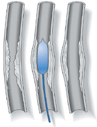
Angioplasty is a medical procedure that opens upblocked or narrowed blood vessels without surgery. An interventional radiologist, a doctor specially trained in minimally invasive, targeted treatments, performs this procedure. During the angioplasty, the interventional radiologist inserts a very small balloon attached to a thin tube (a catheter) into a blood vessel through a very small incision in the skin, about the size of a pencil tip. The catheter is threaded under X-ray guidance to the site of the blocked artery. When the balloon is in the area of the blockage, it is inflated to open the artery, improving blood flow through the area.

Why do I need angioplasty?

The most common reason for angioplasty is to relieve a blockage of an artery caused by atherosclerosis (hardening of the arteries). Atherosclerosis is a gradual process in which cholesterol and scar tissue build up inside the artery, forming a substance called “plaque” that clogs the artery. Arteries are like tubes; they carry blood and oxygen to the tissue in your body. When an artery becomes narrowed or blocked, the tissue supplied by that artery does not get enough oxygen.
The symptoms you feel depend on which artery is blocked. For example, a blocked artery in the legs may cause pain when you walk or even when you are resting in bed. A blocked artery to a kidney may cause high blood pressure. Some blockages are best treated with surgery, while others are best treated with angioplasty.
A member of the interventional radiology team – the doctor, nurse or technologist – will talk with you about the procedure in detail and answer any questions you have. The angioplasty procedure usually takes one to two hours to complete. In some cases, it may take longer.

What is an angioplasty like? Will it hurt?

If you have not had an angiogram (an X-ray examination of your arteries), the interventional radiologist will perform that procedure before proceeding with your angioplasty. An angioplasty has three major steps: placement of the angioplasty catheter into the blocked artery, inflation of the balloon to open the blocked artery, and removal of the catheter. Local anesthetic is used so that you will only feel some pressure during the procedure.

How do I prepare for the procedure?

If you are already a patient in the hospital, your nurses and doctors will give you instructions on how to prepare for your angioplasty. If you are being admitted to the hospital on the morning of your angioplasty, or if you are having your procedure done as an outpatient, follow these instructions, unless your doctor specifies otherwise:

Eating

Do not eat any solid food after midnight on the night before your procedure. You may drink clear fluids.

Medication

Most people can continue to take their prescribed medicines. If you are a diabetic and take insulin, ask your doctor about modifying your insulin dose for the day of your procedure. If you are taking the oral anti-diabetic medicine glucophage (Metformin), you will need to discontinue use for up to 48 hours prior to the procedure and 48 hours following the procedure. Consult with your doctor about blood sugar control during this period. If you take a blood thinner such as Coumadin, you must tell your doctor so that it can be stopped. Bring all your medications with you.

Allergies

If you are allergic to contrast (X-ray dye) or iodine, let your doctor know as soon as possible. If possible, let the interventional radiologist know about your allergy a few days before your angioplasty procedure. Your doctors can then plan to take special precautions during the procedure or prescribe special medications prior to the procedure.

Smoking

Do not smoke for at least 24 hours before your angioplasty. Blood tests are usually done the day before the angioplasty. Before your procedure, you will dress in a hospital gown and an intravenous (IV) line will be placed in one of your veins. You may need to remove your jewelry and any dentures or partials. The IV will be used to give you fluids and medicines during the procedure and will stay in place until after your angioplasty is completed.

Access Menu

What is a biliary catheter?

A biliary catheter is a tube that goes through your skin and liver into your bile ducts to drain your bile. In some cases, the bile drains out of your body into a drainage bag. In other cases, the catheter drains the bile into the bowel and you do not need a bag on the outside. Reasons for needing a biliary catheter include: blockage of the bile ducts, the presence of a hole in the bile ducts, and as preparation for surgery on the bile ducts.

What problems can occur with my biliary catheter?

The main problem that can occur is infection, skin infection around the catheter and bile duct infection. A skin infection can be prevented by taking good care of the skin around the catheter. If a skin infection occurs in spite of good skin care, the infection is usually simple to treat.
A bile duct infection occurs if the catheter gets blocked. The best way to avoid this is to keep your biliary catheter flushed.

How do I take care of the skin around my biliary catheter?

Follow these instructions unless your doctors specify otherwise:
  • Keep the skin around your biliary catheter dry. You can take showers if you cover the area with plastic wrap. Tape the edges of the plastic wrap to your skin so that water cannot get under it. If the area does get wet, dry the skin completely after your shower.
  • Keep the skin around your biliary catheter clean. Clean the area every day or every other day with a cotton swab that has been moistened with peroxide. Always wash your hands before you clean the catheter site.
  • Keep the skin around your biliary catheter covered. After cleaning the skin around the catheter insertion site, cover the area with a clean bandage or dressing. Change the dressing if it gets wet.

What are signs of a skin infection? What should I do for a skin infection?

Signs of a skin infection are redness, soreness, and swelling of the skin around the catheter. If you notice any of these signs, even if they are very mild, you should follow these instructions unless your doctor specifies otherwise:
  • Clean the skin site more often. If you usually clean the skin and change the dressing every other day, start cleaning the skin and changing the dressing every day. If you usually clean the skin and change the dressing once a day, do this twice a day.
  • Apply antibiotic ointment to the skin around the catheter after each time you clean it.
  • If your symptoms of skin infection do improve promptly, keep up the extra care for a total of one week, and then go back to your usual skin care routine.

How do I keep my biliary catheter from becoming blocked, so that I won’t get a bile duct infection?

Sometimes, despite your best efforts, your biliary catheter can become blocked. The following instructions can reduce the risk:
Flush your biliary catheter with sterile saline as your doctor recommends. Usually, this is once a day. Flushing the catheter with saline keeps the inside of the catheter as clean as possible.
If your biliary catheter drains bile into an external drainage bag, rinse the bag out with water every day. It is best to have two bags so that you have one to wear while you are rinsing the other one.
Keep your appointments to have your biliary catheter changed. In most cases, the catheter is changed every two to three months. It is much easier to change the catheter than it is to place the original catheter. This change can usually be done as an outpatient.

How do I know that my biliary catheter is blocked? What are signs of a bile duct infection?

These two problems frequently go together. A sign that your catheter is blocked is leakage of bile around the catheter. Signs that you have bile duct infection are fever and chills.

What should I do if I think my catheter is blocked and/or I think I have a bile duct infection?

Call your interventional radiologist and/or primary doctor immediately. These doctors will arrange for prompt treatment of your problem. In most cases, you will need to have your catheter changed and you may need antibiotic medicine. You may even need to be admitted to the hospital. If your tube is capped off, uncap it and connect it to a drainage bag.

What supplies will I need to take care of my biliary catheter?

You will need to buy:
  • hydrogen peroxide
  • cotton swabs or cotton balls
  • gauze pads
  • surgical tape
  • antibiotic ointment
  • sterile saline
  • syringes
  • needles
  • drainage bags
These are available at drug stores and hospital supply stores. Your doctor will let you know if there are supplies you need that are not on this list.

Do I have to limit my activity?

You will be sore for one to two weeks after your catheter is first inserted. This will limit your activity. After that, you should continue to avoid any activity that causes a pulling sensation or pain around the catheter. There are no special diet instructions.

I know I need to call my doctor if: I have a skin infection that does not go away with care at home, I think my catheter is blocked, or I suspect a bile duct infection. Are there other times I should call my doctor about the catheter?

Yes. Call your doctor immediately if:
  • your catheter becomes dislodged or broken.
  • you have stitches and they become loose.
  • your catheter begins to leak.
  • there is blood in or around your catheter.

Which doctor do I call about my biliary catheter?

Most people who need a biliary catheter have more than one doctor. Your catheter was placed by a specially trained doctor called an interventional radiologist. This specialist works with your other doctors (such as your surgeon, internist, or family doctor) to take care of you once you have a biliary catheter. Your team of doctors may prefer that you contact your interventional radiologist directly if you have a question or problem relating to your catheter. Or, your medical team may prefer that you contact your surgeon, internist, or family doctor first.

Access Menu

What is biliary drainage?

Biliary drainage is a procedure in which a catheter is placed through your skin and into your liver to drain your bile and is performed without surgery. A specially trained doctor, known as an interventional radiologist, performs the procedure in the radiology department. The doctor uses X-ray imaging to help precisely place the catheter.

Why do I need biliary drainage?

The most common reason for biliary drainage is blockage of the bile ducts. The liver makes bile, which aids digestion. Normally, ducts (which are like pipes) carry the bile from the liver into the bowel. When your bile ducts are blocked, the bile backs up in your liver. Signs that your bile ducts are blocked include jaundice (yellow skin color), dark urine, light stools, nausea and poor appetite. Some people experience severe itching.
Biliary drainage can relieve these symptoms by giving the bile a pathway to leave the liver.Biliary drainage may also be needed if a hole forms and bile leaks from the duct. This leakage can cause pain and severe infection. Biliary drainage can stop the leaking and help the hole to heal over.
Biliary drainage may also be needed in preparation for surgery or for some other procedure on your bile ducts, such as removal of a bile duct stone.

How do I prepare for my biliary drainage?

If you are already a patient in the hospital – your nurses and doctors will give you instructions on how to prepare for your biliary drainage.
If you are being admitted to the hospital on the morning of your biliary drainage 0R if you are having your procedure done as an outpatient – follow these instructions unless your doctor specifies otherwise:
Eating. Do not eat any solid food after midnight on the night before your procedure. You may drink clear fluids.
Medication. Most people can continue taking their prescribed medicines. If you are a diabetic and take insulin, ask your doctor about modifying your insulin dose for the day of your procedure. If you take the blood thinner Coumadin, you must tell your doctor so that it can be stopped. Bring all your medications with you.
You will need to have blood work before your biliary drainage. On the day of the procedure, an intravenous (IV) line will be placed in one of your veins and antibiotics will be given. The antibiotics help to prevent infection. The IV will be used to give you other medicines and fluids during the procedure.
Before our biliary drainage begins, a member of the interventional radiology team (doctor, nurse, or technologist) will discuss the procedure with you in detail and answer any questions you might have. The team member will also ask you some general questions about your health and any possible allergies.

What is a biliary drainage procedure like? Does it hurt?

Before the procedure starts, pain medication will be given to you through your IV. Additionally, your interventional radiologist will use local anesthetic to numb the skin and deeper tissues in the area where the catheter will be placed. After that, you will still feel some pressure during the procedure.
Biliary drainage has three major steps: placement of a needle into a bile duct, placement of a guide wire farther into the duct, and placement of the drainage catheter over the wire. The entire procedure usually lasts about two hours, but it may take longer. It is not possible to know in advance exactly how much time your biliary drainage will require.

How do I keep my biliary catheter from becoming blocked, so that I won’t get a bile duct infection?

Sometimes, despite your best efforts, your biliary catheter can become blocked. The following instructions can reduce the risk:

What happens after my biliary drainage?

After the procedure is over, you will go from the radiology department to your hospital room. Your nursing staff will observe you. They will let you know when you can eat and how long you need to stay in bed. Because everyone is different, it is not possible to predict how many days you will need to stay in the hospital.
If you had symptoms of bile duct blockage (such as jaundice) before your biliary drainage catheter was placed, you will notice those symptoms gradually disappear. You will be sore for seven to 10 days after your catheter is inserted.
The biliary drainage catheter is small, about the same size as IV tubing. At first, it will be connected to a drainage bag and your bile will drain into the bag. In many cases, the drainage bag will not be needed after a few days and the catheter will be capped off.

How long will I need the drainage catheter?

How long you will need your catheter depends on why you need your catheter. If the catheter is to be placed to relieve blockage of the bile ducts, you will need the catheter as long as the blockage is present. Your bile ducts can be blocked by stones, infection, scar tissue or tumor. Some patients need their biliary drainage catheter for the rest of their lives. If your catheter is to be placed because you have a hole in your bile duct, you will need the catheter until the hole has healed. If your catheter is to be placed in preparation for bile duct surgery, you will need it until after your surgery. Your doctors will discuss with you how long you are likely to need a biliary drainage catheter.

What are the risks of biliary drainage?

Biliary drainage is safer than surgery, but complications can occur. The two most frequent complications are bleeding and infection. That is why you need to stay in the hospital after the catheter is placed. Because everyone is different, there may be risks associated with your biliary drainage that are not mentioned here. A member of your interventional radiology team will discuss the risks of your biliary drainage procedure with you in detail before the procedure starts.

What are the benefits of biliary drainage?

If your bile ducts are blocked, the biliary drainage catheter will relieve your symptoms, such as jaundice and itching. Before this drainage procedure was developed, patients with blocked bile ducts had to undergo surgery to drain the bile.
In some cases, the catheter can help your doctors eliminate the source of the blockage. For example, if your bile ducts are blocked with stones, your interventional radiologist may be able to remove the stones through the catheter tract without surgery. If your bile ducts are blocked with scar tissue, your interventional radiologist may be able to use instruments through the catheter tract to enlarge the duct in the area of scarring. In some cases, a permanent stent can be placed in the duct to hold it open. Your doctors will talk to you about the best way to manage the cause of your blocked bile ducts.

Access Menu

Our video about Venous Access

What is a Central Venous Access Catheter or CVAC?

A CVAC is a tube that is inserted beneath your skin so there is a simple, pain-free way for doctors or nurses to give you nutrients, or to draw your blood. When you have a CVAC, you are spared the irritation and discomfort of repeated needle injections.
Doctors recommend CVACs for patients who regularly undergo dialysis or chemotherapy treatments, or for patients who frequently receive antibiotic or antifungal infusions or nutritional supplements.
More than 3.4 million CVACs are placed each year, and doctors increasingly recommend their use. There are several types of CVACs, including tunneled catheters (you may hear them called Hickman or Broviac catheters), peripherally-inserted central catheters (sometimes called PICC lines or long lines), dialysis catheters and implantable ports. Your doctor can explain which one is best for you.

What happens when I get a CVAC? Will it hurt?

An interventional radiologist will do the CVAC procedure. You will receive medication to help you relax, and the area will be numbed. A needle is inserted into the skin, creating a small tunnel. The CVAC is then placed in the tunnel with the tip coming to rest in a large vein.You may feel some pressure and slight discomfort during the procedure, but it is not very painful.

How do I prepare for the procedure?

If you already are a patient in the hospital, your nurses and doctors will give you instructions on how to prepare for your CVAC.
If you are being admitted to the hospital on the morning of your CVAC procedure, or if you are having your procedure done as an outpatient, follow these instructions unless your doctor specifies otherwise:

Eating

Do not eat solid food after midnight on the night before your procedure. You may drink clear liquids.

Medication

Most people can continue to take their prescribed medication. If you are a diabetic and take insulin, ask your doctor about modifying your insulin dose for the day of your procedure. If you take a blood thinner, such as Coumadin, you must tell your doctor so that it can be stopped. Bring all your medications with you.

Are there alternatives to CVACs?

The alternative is to continue receiving medication or drawing blood using an IV or needle each time you undergo a procedure. If your doctor recommends a CVAC, its with your best interest in mind. A CVAC will make your frequent treatments more comfortable.

Can I shower after the CVAC procedure?

Most doctors recommend that showering be avoided until the incision heals.

How long will I need to have the CVAC?

It depends on your medical condition. A CVAC may be needed for periods from a few weeks or months to several years. Your doctor can give you more information.

Are there any risks related to a CVAC?

Placement of a CVAC is a safe procedure. The most common complications are infections, blockage of the CVAC and, occasionally, clotting of the blood. All can be resolved with appropriate medical care.

How can I avoid infections related to my CVAC?

To avoid infections, follow these instructions unless your doctor specifies otherwise:
  • Keep the skin around your CVAC dry. After the incision heals, you can take showers, but cover the area with plastic wrap. Tape the edges of the plastic wrap to your skin so that water cannot get in. If the area does get wet, dry the skin completely after your shower.
  • Keep the skin around your CVAC clean. Clean the area every day or every other day with a cotton swab that has been moistened with peroxide. Always wash your hands before you clean the CVAC site.
  • Keep the skin around your CVAC covered. After cleaning the skin around the CVAC site, cover the area with a clean bandage or dressing. Change the dressing if it gets wet.

Do I always need a dressing on my CVAC?

It is important to keep your CVAC very clean. A dressing is usually recommended.

What are the signs of CVAC infection?

Signs of an infection are redness, soreness and swelling of the skin around the CVAC. If you notice any of these signs, even if they are very mild, follow these instructions unless your doctor specifies otherwise:
  • Clean the skin around your CVAC more often. For example, if you usually clean the skin and change the dressing every other day, start cleaning the skin and changing the dressing daily. If you usually clean the skin and change the dressing once a day, do this twice a day.
  • Apply antibiotic ointment to the skin around the CVAC each time you clean it.
  • If your symptoms of infection improve promptly, keep up the extra care for one week. Then go back to your usual CVAC care routine. If the symptoms continue or you have questions or concerns, call your doctor.

How can I avoid blockage of my CVAC?

Sometimes, despite your best efforts, your CVAC can become blocked. To reduce the risk, flush your CVAC with sterile saline once a day. Flushing the CVAC with saline keeps the inside as clean as possible.

How do I know that my CVAC is blocked?

A sign that your CVAC is blocked is leakage of fluid near its opening or feeling resistance when you flush it. If you feel resistance when flushing, do not continue and call your doctor immediately.

What should I do if I think my CVAC is blocked or an infection does not clear up with extra care?

Call the interventional radiologist who placed your CVAC and/or your primary care doctor immediately. The doctor will arrange for proper treatment of your problem. In some cases, you will need to have your CVAC changed, and you may need antibiotic medicine. You may even need to be admitted to the hospital.

I know I need to call my doctor if I have an infection that does not go awaiy with care at home or if I think my CVAC is blocked. Are there other times I should call my doctor about the CVAC?

Yes. Call your doctor immediately if:
  • your CVAC becomes dislodged or broken
  • you have stitches and they become loose
  • your CVAC begins to leak
  • there is blood in or around your CVAC
  • flushing or injecting medication causes pain

Will the CVAC limit my activity?

You will be sore for one to two weeks after your CVAC is first inserted. This will limit your activity. After that, you should continue to avoid any activity that causes a pulling sensation or pain around the CVAC. To avoid unnecessary problems, the catheter that is outside your body should be secured with tape.

What supplies will I need to take care of my CVAC?

Unless your doctor specifies otherwise, you will need to buy:
  • hydrogen peroxide
  • cotton swabs or cotton balls
  • gauze pads
  • surgical tape
  • antibiotic ointment
  • sterile saline
  • syringes
These are available at drug stores and hospital supply stores. Your doctor will let you know if there are supplies you need that are not on this list.

Will the CVAC last as long as I need it?

Yes. Your CVAC should last as long as you need it – anywhere from a few weeks to many years.

What happens when I no longer need my CVAC?

In most situations, the CVAC will be removed. However, if you suffer from chronic, recurring problems, your doctor may choose to leave it in place.

Access Menu

What is Deep Vein Thrombosis (DVT)?

Deep vein thrombosis is the formation of a blood clot in a deep leg vein. It is a very serious condition that can cause death or permanent damage to the leg. In the United States alone, 600,000 people with DVT are admitted to hospitals each year. One in every 100 DVT patients dies.

How does a DVT form?

The deep leg veins are surrounded by powerful muscles that contract to force blood upward against gravity, back to the lungs and heart. One-way valves inside the veins prevent downward back-flow of blood. Blood clots form when the body’s clotting system functions abnormally, and when the circulation of blood slows down due to illness, injury or inactivity.

What are the risk factors for DVT?

Factors that increase your risk for DVT include:
  • Previous DVT or family history of DVT
  • Prolonged immobility, which can be due to stroke, paralysis, prolonged bedrest or sitting, or prolonged air travel (known as “economy class syndrome”)
  • Recent surgery
  • Hormone therapy or oral contraceptives
  • Current or recent pregnancy
  • Trauma and/or orthopedic procedures
  • Varicose veins
  • Congestive heart failure
  • Use of central venous catheters
  • Inflammatory bowel disease
  • Inherited blood clotting (coagulation) abnormalities
  • Obesity
  • Age above 40 years

What are the typical symptoms of DVT?

Common symptoms include:
  • Previous DVT or family history of DVT
  • Prolonged immobility, which can be due to stroke, paralysis, prolonged bedrest or sitting, or prolonged air travel (known as “economy class syndrome”)
  • Swelling of the leg
  • Calf or leg pain or tenderness
  • Leg fatigue
  • Discoloration of the legs
  • Surface veins become more visible

How is a DVT diagnosed?

If you suspect that you may have a DVT, you should immediately go to an emergency room to be evaluated by a physician – it’s a life threatening disease. If your physician agrees that DVT is a possibility, he/she will probably order an ultrasound examination of your leg veins. In some cases, DVT is diagnosed by an Interventional Radiologist using a venogram, which is an X-ray image of your veins – this test allows the physician to see inside your veins and locate a clot that is blocking blood flow. Interventional Radiologists can not only identify the blood clot, but they can also often eliminate the clot and treat underlying problems in the veins that may have caused the blood clot to form.

What problems does DVT cause?

Pulmonary Embolism

When DVT is left untreated, a piece of clot can break off and travel through circulation to the lungs – this is known as pulmonary embolism. The clot can interfere with the lung’s ability to provide oxygen to the body, and when severe, this can cause heart failure and/or death. With early treatment using blood-thinners, people with DVT can reduce their chances of developing a life-threatening pulmonary embolism to less than 1 percent.

Post-thrombotic Syndrome

Post-thrombotic syndrome is an underrecognized but common long-term consequence of DVT. When DVT is treated with blood thinners alone, the blood clot is not actively dissolved. Rather, the blood thinners prevent new clots from forming, but the already present clot remains in the leg. The body may eventually fully or partially dissolve the clot, but the vein valves often become damaged in the meantime. The continued vein blockage and valve damage cause abnormal pooling of blood in the leg, which can cause the post-thrombotic syndrome. Patients with the post-thrombotic syndrome often experience chronic leg pain or heaviness, swelling, difficulty walking, changes in skin color and texture, and in severe cases, skin ulcers (sores). These symptoms may develop within several months or over years. Unfortunately, these problems occur to some degree in as many as 60-70 percent of people with DVT.

How is a DVT treated?

DVT is treated with blood thinners to prevent pulmonary embolism and to prevent further clot formation. In patients with extensive DVT, an additional treatment option is catheter-directed thrombolysis. In patients who cannot receive blood thinners or in whom blood thinners have failed to prevent further clot formation, an additional treatment option is the placement of an inferior vena cava filter.

Catheter-Directed Thrombolysis

Catheter-directed thrombolysis is performed under imaging guidance by Interventional Radiologists. This procedure is designed to rapidly break up the clot, restore blood flow within the vein, and potentially preserve valve function to minimize the risk and severity of post-thrombotic syndrome. Using imaging guidance, the Interventional Radiologist inserts a catheter into a leg vein behind the knee and threads it into the vein containing the clot. A clot-dissolving drug is infused through the catheter directly into the clot and repeated over one to two days. The fresher the clot, the faster it dissolves. A venogram is repeated to demonstrate that the clot has dissolved, and to detect any narrowing in the vein that might lead to future clot formation. If vein narrowing is present, the Interventional Radiologist can treat it using balloon angioplasty or by placing a stent.

Vena Cava Filter

In patients in whom catheter-directed thrombolysis and blood thinners are not medically appropriate, an Interventional Radiologist can insert a small filtering device into the large vein that drains the legs (the inferior vena cava) under imaging guidance. This vena cava “filter” functions like a catcher’s mitt to capture blood clots that break off.

Access Menu

What is a nephrostomy?

A nephrostomy is a procedure in which a catheter is placed through your skin and into your kidney to drain your urine. A specially trained doctor, known as an interventional radiologist, performs this procedure in the radiology department. This doctor uses X-ray imaging to help guide the catheter into exactly the right place to drain your urine. Nephrostomy drainage is performed instead of surgery.

Why do I need a nephrostomy?

The most common reason for a nephrostomy is blockage of the ureter. The kidney makes urine, which drains down the ureter from the kidney to the bladder. When your ureter is blocked, the urine backs up into your kidney. Signs of ureter blockage include pain and fever, but some people have no symptoms. Even if you have no symptoms, a blocked ureter needs treatment because if urine cannot drain out of the kidney, the kidney may stop working. Nephrostomy drainage can relieve the symptoms of ureter blockage and keep the kidney working by giving the urine a way to leave the kidney.
Another reason to need a nephrostomy is if a hole forms in the ureter or bladder, allowing urine to leak into other parts of your body. This leakage can cause pain and severe infection. Nephrostomy drainage can stop the leaking and help the hole heal.
A third reason to need a nephrostomy is to help prepare you for surgery or for some other procedure on your kidney or ureter, such as removal of a large kidney stone.

How do I prepare for my nephrostomy drainage procedure?

If you are already a patient in the hospital – your nurses and doctors will give you instructions on how to prepare for your nephrostomy drainage.
If you are being admitted to the hospital on the morning of your nephrostomy drainage – follow these instructions unless your doctor specifies otherwise:
Eating. Do not eat any solid food after midnight on the night before your procedure. You may drink clear fluids.
Medication. Most people can continue to take their prescribed medicines. If you are a diabetic and take insulin, ask your doctor about modifying your insulin dose for the day of your procedure. If you take the blood thinner Coumadin, you must tell your doctor so that it can be stopped. Bring all your medications with you.
Everybody having a nephrostomy drainage will have blood tests done close to the day of the procedure. On the day of the procedure, an intravenous (IV) line will be placed into one of your veins and antibiotics will be given to you through it. The antibiotics help to prevent infection. The IV will be used to give you other medicines and fluids during the procedure. It will stay in place until after your nephrostomy drainage is complete. Before your nephrostomy drainage begins, a member of the interventional radiology team (doctor, nurse, or technologist) will talk with you about the procedure in detail and answer any questions you may have.

What is nephrostomy drainage like? Does it hurt?

Before the procedure starts, pain medication will be given to you through your IV. Additionally, your interventional radiologist will use local anesthetic to numb the skin and deeper tissues in the area of your back where the catheter will be placed. After that, you will only feel some pressure during the procedure.
Nephrostomy drainage has three major steps: placement of a needle into the kidney, placement of a guide wire farther into the kidney, and placement of the drainage catheter. The procedure usually lasts about one hour, but it is not possible to know exactly how much time your procedure will require.

What happens after the nephrostomy drainage?

After the procedure is over, you will go to your hospital room. Your nursing staff will observe you to make sure you are all right. They will let you know when you can eat and how long you need to stay in bed. Because everyone is different, it is not possible to predict how many days you will need to stay in the hospital.
If you had symptoms of ureter blockage before your nephrostomy drainage catheter was placed, you will notice those symptoms gradually going away. You will be sore for seven to 10 days after your catheter is inserted.
The nephrostomy drainage catheter is about the same size as IV tubing or a bit smaller. The catheter will be connected to a drainage bag and your urine will drain out of your body into the bag. In some cases, the drainage bag will not be needed after a few days and the catheter will be capped off.

How long will I need the drainage catheter?

It depends on why you need your catheter. If the catheter is to be placed to relieve blockage of the ureter you will need the catheter as long as the blockage is present. Your ureter can be blocked by stones, infection, scar tissue, or tumor. Some patients need their nephrostomy drainage catheter for the rest of their lives. If your catheter is to be placed because you have a hole in your ureter, you will need the catheter until the hole has healed. If your catheter is to be placed in preparation for surgery, or another procedureon your kidney or ureter, you will need the catheter until afterwards. Your doctors will discuss with you how long you are likely to need a nephrostomy drainage catheter.

What are the risks of nephrostomy drainage?

Nephrostomy drainage is safe, but complications can occur. The two most frequent complications are bleeding and infection. That is why you need to stay in the hospital after the catheter is placed. Because everyone is different, there may be risks associated with your nephrostomy drainage that are not mentioned here. A member of your interventional radiology team will discuss the risks of your nephrostomy drainage procedure with you in detail before the procedure starts.

What are the benefits of a nephrostomy drainage?

If your ureter is blocked, the nephrostomy drainage catheter will relieve your symptoms, such as pain, fever or chills. Before this drainage procedure was developed, patients with blocked ureters had to undergo surgery to drain the urine.
In some cases, the catheter can help your doctors eliminate the source of the blockage. For example, if your ureter is blocked with stones, your doctors may be able to remove the stones through the catheter tract without surgery. If your ureter is blocked with scar tissue, your doctor may be able to use instruments through the catheter tract to enlarge the ureter in the area of scarring. Your doctor will talk to you about the best way to manage the cause of your blocked ureter.
If you have a hole in one of your ureters, the catheter will drain the urine and help to prevent serious infection while the hole heals. In most cases, this makes surgery to close the hole unnecessary.

Access Menu

What is a nephrostomy catheter?

A nephrostomy catheter goes through your skin into your kidney to drain your urine. In some cases, the urine drains out of your body into a drainage bag. In other cases, the catheter drains the urine into the bladder and there is no bag on the outside. Reasons to need a nephrostomy catheter include: blockage of the ureter (the structure that normally carries urine from the kidney to the bladder), presence of a hole in the ureter, and preparation for surgery or other procedures on the kidney and ureter.

What problems can occur with a nephrostomy catheter?

The main problem that can occur is infection. Your nephrostomy catheter can cause two types of infection: a skin infection and a kidney infection. A skin infection can be prevented by taking good care of the skin around the catheter. If a skin infection occurs in spite of good skin care, it is usually simple to treat.
A kidney infection occurs if the catheter gets blocked. A kidney infection is more serious than a skin infection, but it can be avoided. The best way to avoid a kidney infection is to have your catheter changed for a new one regularly. If your catheter gets blocked and your kidney gets infected in spite of good care, your doctors can treat the problem by changing the catheter and giving you antibiotics.

How do I take care of the skin around my nephrostomy catheter?

Because the catheter is on your back, you will need help. Follow these instructions unless your doctor specifies otherwise:
  • Keep the skin around your nephrostomy catheter dry. You can take showers, but cover the area with plastic wrap. Tape the edges of the plastic wrap to your skin so that water cannot get under it. If the area does get wet, dry the skin completely after your shower.
  • Keep the skin around your nephrostomy catheter clean. Clean the area every day or every other day with a cotton swab that has been moistened with peroxide. Always wash your hands before you clean the catheter site.
  • Keep the skin around your nephrostomy catheter covered. After cleaning the skin of the catheter insertion site, cover the area with a clean bandage or dressing. Change the dressing if it gets wet.

What are signs of a skin infection? What should I do for a skin infection?

Signs of a skin infection are redness, soreness, and swelling of the skin around the catheter. If you notice any of these signs, even if they are very mild, you should follow these instructions unless your doctor specifies otherwise:
  • Clean the skin site more often. If you usually clean the skin and change the dressing every other day, start doing this every day. If you usually clean the skin and change the dressing once a day, do this twice a day.
  • Apply antibiotic ointment to the skin around the catheter after each time you clean it.
  • If your symptoms of skin infection do not improve within 48 hours, or if they worsen despite the extra care, call your doctor.
  • If your symptoms of skin infection do improve promptly, keep up the extra care for a total of one week, and then go back to your usual skin care routine.

How do I keep my nephrostomy catheter from getting blocked so that I won’t get a kidney infection?

You cannot completely avoid the possibility that your nephrostomy catheter will get blocked. However, the instructions listed below can minimize the risk:
  • If your nephrostomy catheter drains urine into an external drainage bag, rinse the bag out with water every day. It is best to have two bags so that you can wear one while you are rinsing the other one.
  • Apply antibiotic ointment to the skin around the catheter after each time you clean it.
  • Keep your appointments to have your nephrostomy catheter changed. In most cases, the catheter is changed every two to three months. Changing the catheter helps to prevent catheter blockage. The longer the catheter is in, the more likely it is to get blocked. It is much easier to change the catheter than it is to place the original catheter. This change can usually be done as an outpatient.

What are signs that my nephrostomy catheter is blocked? What are signs of kidney infection?

These two problems frequently go together. Your nephrostomy catheter may get blocked and cause kidney infection in spite of good care. Signs that your catheter is blocked are back pain and leakage of urine around the catheter onto your skin and catheter dressing. Signs that you have a kidney infection are pain, fever, and chills.

What should I do if I think my catheter is blocked and/or I think I have a kidney infection?

Call your interventional radiologist and/or primary doctor immediately. These doctors will arrange for prompt treatment of your problem. In most cases, you will need to have your catheter changed and you will need antibiotic medicine. You may need to be admitted to the hospital. If your tube is capped off, uncap it and connect it to a drainage bag.

What supplies will I need to take care of my nephrostomy catheter?

You will need to buy:
  • cotton swabs or cotton balls
  • hydrogen peroxide
  • gauze pads
  • surgical tape
  • antibiotic ointment
  • drainage bags
These things are available at drug stores and hospital supply stores. Your doctor will let you know if there are supplies you need that are not on this list.

Do I have to limit my activity?

You will be sore for one to two weeks after the catheter is first inserted. This will limit your activity. After that, you should continue to avoid any activity that causes a pulling sensation or pain around the catheter or kinking of the catheter.

I know I need to call my doctor if: I have a skin infection that does not go away with care at home, I think my catheter is blocked, or I suspect I have a kidney infection. Are there other times I should call my doctor about the catheter?

Yes. Call your doctor immediately if:
  • your catheter becomes dislodged or broken.
  • you have stitches and they become loose.
  • your catheter begins to leak.
  • there is blood in or around your catheter.

Which doctor do I call about my nephrostomy catheter?

Most people who need a nephrostomy catheter have more than one doctor, and it can be difficult to know which doctor to call when you have questions about or a problem with your catheter. Your catheter was placed by a specially trained doctor called an interventional radiologist. This specialist works with your other doctors (such as your surgeon, internist, or family doctor) to take care of you once you have a nephrostomy catheter. Your team of doctors may prefer that you contact your interventional radiologist directly if you have a question or problem relating to your catheter. Or, your medical team may prefer that you contact your surgeon, internist, or family doctor first. Ask your doctors whom you should call when you need advice or help with catheter care.

Access Menu

What is gastrostomy?

A gastrostomy is performed to insert a tube directly into the stomach or small intestine of a person who cannot take food or medicine by mouth. Gastrostomy can be done surgically, or through an interventional radiology (IR) technique called “percutaneous gastrostomy,” which requires only a tiny incision in the skin. Percutaneous gastrostomy can be performed safely in adults or children. Generally, this is an outpatient procedure or may involve a short hospital stay.

Why is a gastrostomy tube placed?

Gastrostomy tube placement is performed for a variety of reasons. Some gastrostomies are performed to provide a route for feeding in people who are unable to eat and swallow normally. Gastrostomies may also be advised for people with chronic bowel obstruction who cannot tolerate a nasogastric tube (a tube inserted through the nose into the stomach). Your physician will explain the reason for your gastrostomy.

How can I arrange for an interventional radiologist to perform my percutaneous gastrostomy?

Evaluation may require blood tests and a consultation with a clinical nutritionist who can explain all about percutaneous gastrostomy feedings. You will be informed about the benefits and risks of having the procedure, and any questions you may have will be answered.

How should I prepare for a percutaneous gastrostomy?

You must not eat, drink or receive nasogastric tube feedings for at least six hours before the percutaneous gastrostomy procedure. However, you may take routine medications by mouth with small sips of liquid, or medications may be given through a nasogastric tube. More detailed instructions, based on your individual needs, will be given to you by your interventional radiologist when your procedure is scheduled.

What happens on the day of the procedure?

You will change into a hospital gown and go to the interventional radiology suite where the procedure will be performed. Usually, the procedure is performed using a mild sedative and local anesthesia. In rare cases, general anesthesia is required and an anesthesiologis will work with the interventional radiologist to monitor you during the procedure.
Once you are in the interventional radiology suite, the following steps will be taken to perform the gastrostomy procedure:
  • An intravenous line will be placed in your arm to deliver the anesthetic or sedative, analgesics (medication to prevent pain), and an antibiotic (medication to prevent infection), if needed.
  • An ultrasound scan of the stomach area may be performed to assist your interventional radiologist in selecting the correct placement of the gastrostomy tube – usually below the rib cage.
  • Your stomach will be inflated with air. The air is usually delivered to the stomach through a nasogastric tube that is inserted through the nose and into the stomach.
  • The skin over the stomach area will be cleansed with an antiseptic solution and draped with sterile covers. A local anesthetic will be injected in the skin to numb the area where the tube will be placed. In some situations, the stomach is first fastened to the abdominal wall by inserting small devices through the skin. These are later removed.
  • The tube will be inserted by the interventional radiologist through a very tiny incision in the skin – about five millimeters long (approximately 1/4 inch).
  • Typically, percutaneous gastrostomy is performed in less than an hour. The procedure is painless, but there might be some mild discomfort related to the temporary expansion of your stomach and placement of the gastrostomy tube. Immediately after the procedure, you will be transferred to a recovery unit where you will be monitored.

What will happen after the gastrostomy? Can I go home?

Most adult patients and some older children may be discharged to go home the same day as the procedure, and arrangements are generally made for a home care nurse to visit during the first 24 to 48 hours. Most young children will be admitted to the hospital following gastrostomy. If the interventional radiologist has used fastening devices to insert the tube, you will be instructed to return to your interventional radiologist at five to seven days for a simple outpatient removal of these tiny fasteners. You will be given written instructions before you go home about gastrostomy tube care, feeding, warning signs of possible problems and telephone numbers to call in case you have questions.

Feeding guidelines for gastrostomy tubes

The following guidelines may be helpful for your feedings after percutaneous gastrostomy, although your interventional radiologist and clinical nutritionist may have more specific recommendations for you.
  • Maintain an upright position during your feeding and for 30 to 60 minutes after feeding. Lying on your right side also may help the food or liquid formula enter your body through the tube.
  • Make sure the food or formula is not too cold. It is helpful to warm formula to room temperature before administering.
  • Make sure the formula has not expired or has not been open more than 24 to 48 hours. To prevent spoilage, keep all open formula in the refrigerator until it is used.
  • Make sure equipment is very clean.
  • Usually, patients are instructed to have water between feedings or immediately following a feeding to prevent dehydration. Also, flushing the tube with water after each feeding will keep it from becoming clogged. The volume of water that can be safely given variesaccording to your weight. Check with your physician, nurse or clinical nutritionist to determine the most appropriate amount of water for you.
  • Medication may also clog tubes. Consult with your physician or pharmacist about your medications if gastrostomy tube clogging becomes a problem.
  • If you have any problems with your gastrostomy, call your interventional radiologist.

Access Menu

What is peripheral vascular disease?

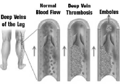
Peripheral vascular disease, or PVD, is a condition in which the arteries that carry blood to the arms or legs become narrowed or clogged, interfering with the normal flow of blood. The most common cause of PVD is atherosclerosis (often called hardening of the arteries). Atherosclerosis is a gradual process in which cholesterol and scar tissue build up, forming a substance called “plaque” that clogs the blood vessels. PVD may also be caused by blood clots.

What are the symptoms of PVD?

PVD Symptoms

  • Leg or hip pain during walking
  • The pain stops when you rest
  • Numbness
  • Tingling
  • Weakness in the legs
  • Burning or aching pain in feet or toes when resting
  • Sore on leg or foot that won’t heal
  • Cold legs or feet
  • Color change in skin of legs or feet
  • Loss of hair on legs
The disease, which affects both men and women, often goes undiagnosed and many people mistakenly think the symptoms are a normal part of aging.
The most common symptom of PVD is called intermittent claudication, a painful cramping in the leg or hip, particularly when walking. Intermittent claudication occurs when there is not enough blood flowing to the leg muscles during exercise. The pain typically goes away when the muscles are given a rest.
Other symptoms may include numbness, tingling or weakness in the leg. In severe cases, you may experience a burning or aching pain in the feet or toes while resting, or develop a sore on the leg or foot that does not heal. People with PVD may also experience a cooling or color change in the skin of the legs or feet, or loss of hair on the legs. In extreme cases, untreated PVD can lead to gangrene, a serious condition that may require amputation of a leg or a foot.
If you have PVD, you are also at higher risk for heart disease and stroke.

Who is at risk for PVD and intermittent claudication?

As many as 10 million people in the U.S. may have PVD. It is estimated that 4 million of those suffer leg pain symptoms. Those who are at highest risk are:
  • over the age of 50,
  • smokers,
  • diabetic,
  • overweight,
  • people who do not exercise, or
  • people with high blood pressure or high cholesterol.

A family history of heart or vascular disease may also put you at higher risk for PVD.

How is PVD diagnosed?

The most common test for PVD is the ankle-brachial index (ABI), a painless exam in which ultrasound is used to measure the ratio of blood pressure in the feet and arms. Based on the results of your ABI, as well as your symptoms and risk factors
for PVD, the physician can decide if further tests are needed. PVD also can be diagnosed noninvasively with an imaging technique called magnetic resonance angiography (MRA), or with computed tomography (CT) angiography.

How can PVD be treated?

The best treatment for PVD depends on a number of factors, including your overall health and the severity of the disease. In some cases, lifestyle changes are enough to halt the progression of PVDand manage the disease. Your physician may prescribe drugs when lifestyle changes are not enough. Procedures that open clogged blood vessels also are used to treat PVD.
Lifestyle changes. Most treatment plans will include a low fat diet and a program of regular exercise. If you are a smoker, it is absolutely essential that you stop the use of all types of tobacco. If decreased blood flow to the legs is causing injury to the feet and toes, a foot care program to prevent sores or infection may be prescribed. This may include referral to a podiatrist.
Medication. Medications that lower cholesterol or control high blood pressure may be prescribed. Medication also is available that has been shown to significantly increase pain-free walking distance and total walking distance in people with intermittent claudication. Other medications that help prevent blood clots or the build-up of plaque in the arteries are available, as well.

What can be done to treat PVD when lifestyle changes and medications are not enough?

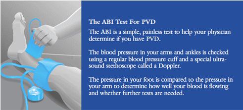
There are a number of ways that physicians can open blood vessels at the site of blockages and restore normal blood flow. In many cases, these procedures can be performed without surgery using modern, interventional radiology techniques. Interventional radiologists are physicians who use tiny tubes called catheters and other miniaturized tools, and X-rays to do these procedures.

Procedures performed by interventional radiologists include:

  • angioplasty – a balloon is inflated to open the blood vessel.
  • thrombolytic therapy – clot-busting drugs are delivered to the site of blockages caused by blood clots.
  • stents – a tiny metal cylinder, or stent, is inserted in the clogged vessel to act like a scaffolding and hold it open.
  • stent-grafts – a stent covered with synthetic fabric is inserted into the blood vessels to bypass diseased arteries.
Sometimes, open surgery is required to remove blockages from arteries or to bypass the clogged area. These procedures are performed by vascular surgeons.

How can I find out if I have PVD?

If you suspect that you may have PVD, it is important that you see your personal physician for an evaluation.